13 Dec 2018
5 min read
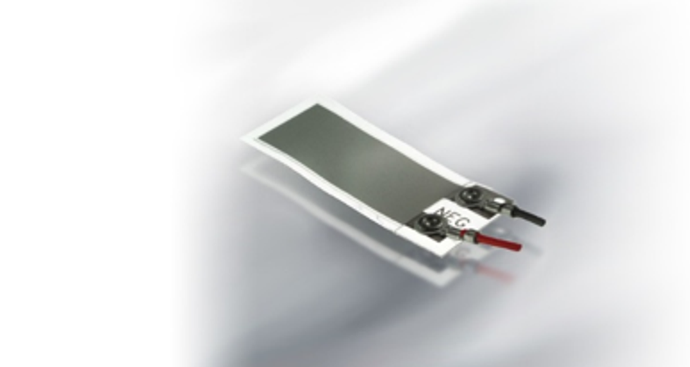
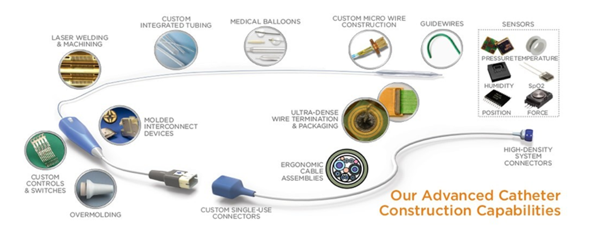
With the ever growing and aging population, patient auto-monitoring systems are becoming more and more popular. Their popularity stems from being both consistent and repeatable in addition to being low cost. Sensor-studded monitoring instruments in this category are also versatile because they can be used both in hospitals and at home. Selecting a sensor can be simple if the application and the parameters that need to be monitored are clearly understood. The most complicated sensors are implantables, followed by sensors used in catheters (through incision) and sensors used in body cavities, sensors that are external but come in contact with body fluids and sensors for external applications. IMPLANTABLE SENSORS Implantable sensors need to be small, lightweight, and compatible with body mass as well as require very little power to operate. Most importantly, they must not decay over time. Since they are Class III medical devices, they automatically require FDA approval. Implantable sensors typically require two to four years for development and implementation before moving on to production. Generally, they are more expensive and require a specialist to surgically implant them. The power requirement is one of the major challenges for implantable sensors. Sensors that can function with no power are ideal, but these are few and rare in the market. Piezoelectric polymer sensors are well suited for vibration detection since they are small, reliable, durable, and require no power. Such sensors can be used in pacemakers that monitor activities of the patient. This Piezo sensor is in the shape of a tiny cantilever beam with weight attached on one end that flops with body movement. Every time the patient moves, the sensor generates a signal. Using a pacemaker as an example, the pacemaker then receives this signal and makes the heart beat at the desired pace. The sensor can differentiate between various activities such as walking, running, or other physical activities. For instance, if the patient is resting, the signal will be zero and the pacemaker will make the heart beat at a minimal rate. In this way, the sensor signal is proportional to the level of activity. A miniature Piezo film vibration sensor is 15/100 of an inch in length including the pacemaker which houses it. Implanted sensors can also be powered by external sources. For example, a Radio Frequency (RF) energy wand when placed near a sensor located inside the body will power the sensor up. The sensor will then record patient measurements, transmit the data back to the wand via RF link, and return to hibernation. Another example of using an implanted sensor in this way is that of a post abdominal aortic aneurysm procedure where an implanted sensor can monitor the pressure leaks at the surgical location.

SENSORS IN CATHETERS AND BODY CAVITIES The requirements for sensors that can be inserted through an incision – typically at the tip of a catheter – are less critical than those for implantables but will still need FDA approval. Depending on the surgical procedure, these sensors need to function for a few minutes up to a couple of hours and can be powered via external sources. A pair of matched thermistors at the tip of a catheter can be guided to different locations of the heart to measure blood flow. Either they can be heated through the coil or flushed with cold fluid to measure blood flow rates. When flushed with cold fluid, the first sensor gets cooled more than the second because the blood flow warms up the fluid that reaches the second sensor. Since these two temperature sensors are separated by a known distance where the temperature and volume of the fluid is controlled, the blood flow can be calculated by reading the difference of the resistance values of the two sensors. These thermistors do not require external power. Catheter Ablation Sensors These sensors are temporarily inserted through incision. The catheter tip contains a source for RF energy and a force load cell sensor. RF energy, like that used with implantable sensors to send data, is often used in the ablation process to burn out dead tissue. It is critical that the force applied by the catheter tip to the target tissue not exceed maximum values to avoid any possibility of perforating the target tissue. TE's sensing technology holds the promise of providing a triaxial force-sensing system able to measure tissue contact forces in all three dimensions simultaneously.
From Your Site Articles